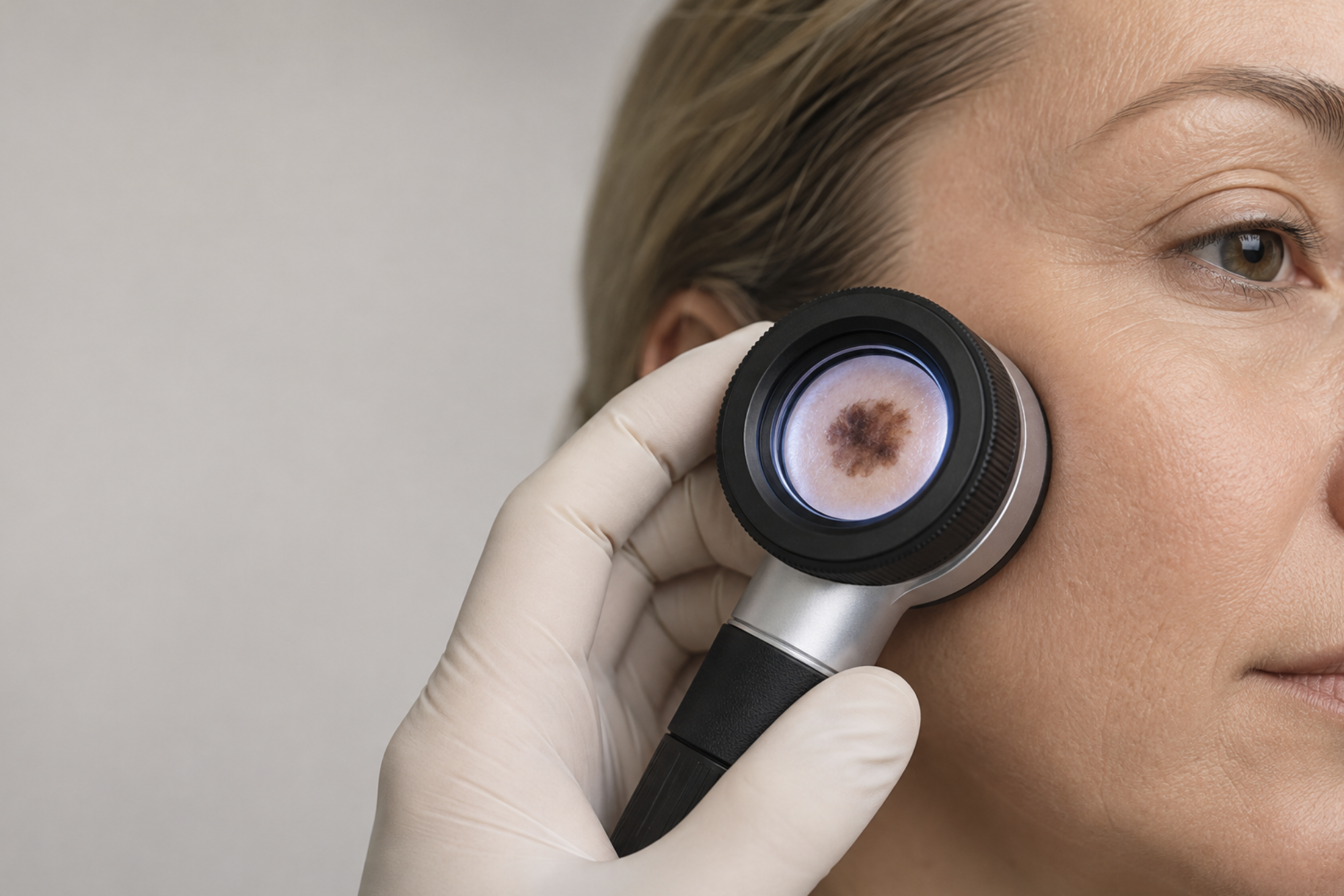
Skin cancer on the face does not always look dramatic. It may appear as a small pearly bump, a rough scaly patch or a sore that repeatedly scabs and bleeds. Because these changes can resemble harmless spots, scars or areas of sun damage, facial skin cancer can sometimes be overlooked in its early stages.
Most suspicious skin lesions will not prove to be cancer; but a new, changing or non-healing area should always be assessed by a medical professional.
Why is skin cancer common on the face?
Ultraviolet radiation from the sun can damage DNA within skin cells. The face, scalp, nose, ears and neck receive considerable cumulative exposure, making them common sites for both basal cell carcinoma and squamous cell carcinoma.
Skin cancer can affect every skin tone. Its colour and appearance may differ between individuals, and cancer should not be ruled out simply because a lesion does not match a photograph seen online.
What does basal cell carcinoma look like?
Basal cell carcinoma, or BCC, is the most common form of skin cancer in the UK. It usually grows slowly and only very rarely spreads elsewhere, but it can damage nearby tissue when left untreated.
A facial BCC may look like:
- A shiny, pearly or translucent bump
- A pink, red, brown or darker patch
- A sore that heals and then returns
- A lesion that bleeds with little provocation
- A scaly area with a raised edge
- A scar-like patch without a history of injury
- A small growth containing visible blood vessels
BCCs can occur around delicate structures such as the nose, eyelids, lips and ears. Early assessment can make treatment and reconstruction more straightforward.
What does squamous cell carcinoma look like?
Squamous cell carcinoma, or SCC, can behave more aggressively than BCC and has a greater potential to spread.
An SCC may appear as:
- A firm or tender lump
- A rapidly growing skin lesion
- A crusted or scaly patch
- A persistent ulcer
- A growth that bleeds
- A raised lesion with a central crater
- A rough area developing on chronically sun-damaged skin
Can melanoma develop on the face?
Yes. Melanoma is less common than BCC and SCC but is more likely to spread.
The ABCDE guide can help identify a suspicious mole:
- A – Asymmetry: one half differs from the other
- B – Border: irregular, blurred or notched edges
- C – Colour: uneven or multiple colours
- D – Diameter: increasing size, although melanoma can be small
- E – Evolution: any change in size, shape, colour, sensation or behaviour
It’s important to remember that some melanomas are pink or relatively colourless, so darkness is not essential.
When should a facial lesion be checked?
Arrange an assessment if a lesion:
- Has not healed after several weeks
- Repeatedly scabs or bleeds
- Is growing or changing
- Becomes painful, itchy or tender
- Looks different from your other marks
- Appears on a previously treated skin-cancer site
- Concerns you in any way
What happens during assessment?
Your clinician will ask how long the lesion has been present and whether it has changed. It will be examined directly and may also be assessed using dermoscopy, which provides a magnified view of structures beneath the skin’s surface.
Depending on its appearance, the next step may be monitoring, biopsy or complete removal. Tissue is examined under a microscope to establish the diagnosis.
Treatment depends on the cancer type, size, depth and location. Surgery may involve standard excision, Mohs micrographic surgery or another carefully selected approach. When cancer occurs on the face, planning must consider both complete removal and reconstruction.
Mr Ross Elledge is a fellowship-trained Consultant Oral and Maxillofacial Surgeon with specialist experience in facial skin cancer diagnosis, treatment and reconstruction. If you’re worried about a facial lesion or have recently received a diagnosis and would like to discuss reconstruction options, get in touch.